Tooth Decay

Early tooth decay

Early tooth decay is usually painless and the tooth surface seems intact when examined by the naked eye. Tooth decay at the adjacent tooth surfaces is even more difficult to be detected and they often need to be confirmed by radiographs.

Photograph of the longitudinal section of a tooth crown showing initial decay.

Treatment:

The dentist may apply high concentration topical fluoride such that early tooth decay lesions can be repaired.

Animation showing the longitudinal section of a tooth at its initial stage of tooth decay.  An appropriate amount of fluoride has been applied to the decay resulting in the remineralization of the decayed portion.

Tooth decay spreads into dentine

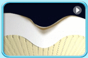
A cavity may appear on the tooth and discomfort may be felt on eating.

Animation of the longitudinal section of a tooth crown with decay started in enamel and then all the way to dentine leading to apparent cavity.

Treatment:

  • A filling can be placed if the tooth decay lesion is relatively small and the structure of the tooth remains strong.
  • When tooth decay lesion is wide-spread, it may render the remaining tooth structure weak. A crown may have to be made for protection.

Tooth decay spreads into pulp

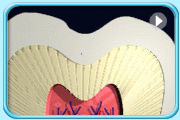
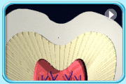
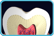
At this stage, cavity appears on the tooth and causes severe pain. The pulp tissues are infected by the bacteria and may become necrotic. The bacteria may spread from the pulp to the surrounding periodontal tissues via the apex of the tooth, leading to inflammation or even the formation of abscess.

Animation of the longitudinal section of a tooth crown showing that the decay starts from enamel and then all the way to the dentine leading to pulp infection and inflammation.

Treatment: